Brain Imaging
Frequently Asked Questions (FAQs)
What is MRI and fMRI? Does MRI/fMRI use radiation?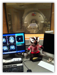
No. MRI uses a powerful magnetic field, radio frequency pulses, and a computer to produce detailed pictures of organs, soft tissues, bone and virtually all other internal body structures. MRI does not use ionizing radiation (x-rays, CT, CAT).
Functional magnetic resonance imaging (fMRI) is a relatively new procedure that uses MR imaging to measure the tiny metabolic changes that take place in an active
part of the brain. In our study, we look at blood-oxygen-level dependent (BOLD) imaging to observe areas of the brain that are active (or have high levels of oxygen).
Why can’t my child have any metal in/on their body?
An MRI machine uses a magnetic field to detect differences in tissue types (structural scans) and changes in blood oxygen levels (BOLD of fMRI). The magnetic field is very strong and could pull certain metal objects (especially objects that contain a lot of iron) away from the body or even heat up the metal. For safety reasons, we ask subjects to remove any jewelry, watches, cell phones, and wallets before entering the scanner.
We also perform safety screenings to determine eligibility for the imaging portion of this study. Subjects who have permanent metal in their body (such as pins, rods, heart pacemakers, cochlear implants, braces, etc.) would not be allowed in an MRI scanner.
For scientific reasons, certain metals could also disrupt the images we are taking of the head. Although these may not be safety concerns, we would ask participants to make sure that they do not have these things, in order to get good quality images. Common examples include:
- Hair ties or bobby pins with metal on them
- Permanent retainers
- Metallic eye shadow/liner
- Certain hair products that include metal or metallic properties
Do you inject my child with contrast agents?
No. By using BOLD imaging, we do not need to inject your child with any contrast agents. Changes in oxygen levels occur naturally in the body and the magnetic field of the MRI scanner can detect these changes (remember, hemoglobin is an iron-based molecule, which is why our blood is red).
Will I get to enter the scanner room with my child?
For this study, parents will not be allowed into the scanner room. Individuals allowed into the scanner room must first complete an fMRI metal screening form with a member of our research team. All paperwork then has to go through the Waisman Center Brain Imaging Department.
Although parents will not be allowed into the scanner room, parents will be allowed into the control room next to the scanner room at the start of the scan while we prepare your child for their brain scan. You will be able to view your child through a large window that connects the two rooms. Once your child is comfortable laying in the scanner, we will have you wait out in the lobby.
Does my child get a picture of their brain?
We’re sorry, no. The Waisman Center has a strict policy regarding viewing brain images. All images must first go through a radiologist who checks for clinical findings (for more information related to unexpected discoveries see final question).
Additionally, this study currently does not have approval from the Institutional Review Board (IRB) to distribute brain images to participants even after going through a radiologist and completing the study.
The consent form states that the research scanner we are using is not the same quality as the ones in a hospital and therefore should not be used for diagnostic purposes. What makes it different?
MRI scanners can and do generate a variety of different ways of taking pictures, called “pulse sequences”. The pulse sequences we choose are optimized for our research questions, which center on math learning and development in certain regions of the brain. For example, we perform a T1 weighted scan (structural scan) of the entire brain within just 8 minutes. When doing this quick scan, we are not looking for abnormalities.
On the other hand, if you were using a hospital MRI, a physician might select a smaller region of the brain or might take a longer scan to get a higher resolution image of the brain. Similarly, a physician may even use contrast imaging to identify certain things, while we do not.
Unexpected findings – uncertain significance vs clear significance?
Remember that *all* scans are reviewed by a member of the radiology staff, who is a physician trained to look for any unusual findings and to make a determination of whether or those findings are clinically significant or not.
Uncertain clinical significance means the brain image shows something unusual in the brain, but we do not know if/how it may affect your child’s health. Treatment may not be appropriate or possible. You will not be informed of uncertain findings.
Clear clinical significance means the brain image shows something unusual that is well known, may be treatable, and risks of not treating the problem are known. You will be informed of certain findings. You may also choose to have your primary physician informed by providing his/her name and location on the parent consent form.